Microtubule Staining: Techniques and Applications


Intro
Microtubule staining is a cornerstone in cellular biology, acting as a window into the microcosm of cell structures. Often overlooked, these cytoskeletal components are fundamental to cell shape, motility, and division. The examination of microtubules extends far beyond mere observation; it dives into the intricate relationships that govern cellular activities. As researchers grapple with questions surrounding disease mechanisms and cellular interactions, understanding microtubules through staining becomes increasingly vital.
This piece aims to dissect the various methodologies employed in microtubule staining. We'll explore the tools and technologies that have transformed the field, shedding light on their applications in scientific inquiry. Moreover, we'll venture into the advancements that have revolutionized imaging techniques, granting us clearer and more detailed views of these structures. Ultimately, this document serves to synthesize information gathered through years of study, offering a comprehensive overview of microtubule staining and its place in the future of cytoskeletal research.
In following sections, methodologies will be elaborated upon, highlighting the key techniques, tools, and their implications in cellular research. From traditional methods to cutting-edge advancements, the narrative aims to provide insight valuable to students, researchers, educators, and professionals alike.
Prologue to Microtubules
Microtubules are not just an occasional mention in cellular biology; they are fundamental to the architecture and function of eukaryotic cells. The significance of understanding these structures goes beyond mere academic curiosity. They form part of the cytoskeleton, acting like scaffolding that maintains cellular shape and facilitates movement, among other functions. This article endeavors to present a comprehensive exploration of microtubules, spotlighting their staining techniques, applications in various research fields, and groundbreaking advances.
Definition and Structure
Microtubules are cylindrical polymers made primarily of tubulin proteins, characterized by their dynamic nature. They have a diameter of about 25 nanometers, which, despite being minuscule, plays a monumental role in cellular processes. Structurally, they are composed of alpha and beta tubulin dimers that assemble into protofilaments. These protofilaments further align to form the hollow tube. The ability to grow and shrink in response to cellular needs is an incredible feature that allows the cell to adapt rapidly to various stimuli. Their intricacies can be visualized more effectively using staining techniques that highlight the distinct pathways and organizations within the cell.
Role in Cellular Function
The roles that microtubules play are as diverse as they are critical. They act as highways for intracellular transport, enabling the movement of organelles, vesicles, and even proteins. Moreover, during cell division, microtubules form the mitotic spindle, which is vital for the correct segregation of chromosomes. Without microtubules, cells would face substantial challenges in maintaining their integrity and functionality.
In addition, microtubules are involved in more specialized functions, such as the formation of cilia and flagella, which facilitate movement in certain cell types. This versatility makes studying microtubules invaluable for understanding disease mechanisms, as alterations in their function can lead to various disorders, including cancer and neurodegenerative diseases.
Importance in Research
The study of microtubules is a cornerstone of modern cellular biology and biomedicine. Recognizing their structure and function can provide insights into pathophysiological states. Researchers harness staining techniques to visualize these structures, enabling the tracking of their behavior in live cells. With the ongoing advancements in staining technologies, our capability to observe microtubules with greater clarity and resolution has drastically improved.
Research efforts often focus on various applications, from cancer research, which examines how microtubule dynamics might influence tumor growth, to understanding neurodegenerative disorders where microtubule stability is a key concern. The exploration of microtubules may not only help clarify the underlying mechanisms of these conditions but can also pave the way for innovative therapeutic strategies.
Ultimately, the field is a testament to the intricate dance of molecular components where microtubules take center stage.
Staining Techniques Overview
In the study of microtubules, the techniques employed for staining these structures are not just a step in the process; they form the backbone of our understanding of cellular mechanics. This section focuses on the key staining techniques utilized in microtubule studies, examining their effectiveness, advantages, and unique attributes. By analyzing various methods, researchers can select appropriate techniques based on their specific needs, ensuring accuracy and depth in their findings. Thus, each method serves a pivotal role in elucidating the complex dynamics of microtubules, which are essential for numerous cellular processes.
Types of Staining Methods
Staining techniques vary widely, and each serves a particular purpose in visualizing cellular components. Below, we delve into the primary methods utilized to stain microtubules, shedding light on their characteristics and implications for research.
Fluorescent Staining
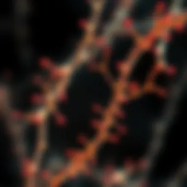
Fluorescent staining is one of the most widely used methods for visualizing microtubules due to its ability to provide real-time insights into cellular structures. The key characteristic here is the use of fluorescent dyes that bind specifically to tubulin, the protein that makes up microtubules. This specificity allows for a precise highlight of microtubule pathways within cells.
A significant benefit of fluorescent staining is its versatility. Various fluorescent dyes, such as Alexa Fluor and FITC, can be utilized depending on the requirements of the experiment. One unique feature of these dyes is their color spectrum, allowing multiple targets to be studied in a single sample through multiplexing. However, one must also consider potential disadvantages, such as photobleaching, which can occur with prolonged exposure to light, leading to loss of fluorescence over time.
Immunofluorescence
Immunofluorescence builds upon the principles of fluorescent staining by specifically targeting antigens in cells. This technique employs antibodies tagged with fluorescent dyes that bind to specific proteins, facilitating a more detailed view of microtubules in the context of cellular function.
The strength of immunofluorescence lies in its specificity and sensitivity. It allows researchers to study not only the location of microtubules but also their interactions with other cellular components. This method is particularly beneficial for exploring how microtubules change during various cellular processes. However, the complexity of using antibodies can lead to variability in results, and the need for extensive controls can be considered a drawback.
Phase Contrast Microscopy
Phase contrast microscopy offers a powerful alternative to staining, providing optical enhancements to enhance visibility without the need for dyes. This technique manipulates light waves to illuminate the phase differences in thick samples, leading to clear images of microtubules in their native environments.
One of the main advantages of phase contrast microscopy is that it allows for live cell imaging. This can provide invaluable insights into dynamic processes, such as cell division. However, the downside is that certain details may not be adequately resolved, which can impede the analysis of fine structural aspects of microtubules.
Electron Microscopy
Electron microscopy, particularly transmission electron microscopy (TEM) and scanning electron microscopy (SEM), takes microtubule imaging to the next level through the use of electrons rather than light. This provides exceptionally high-resolution images that reveal intricate details of microtubule structure.
The exceptional resolution of electron microscopy allows for the visualization of microtubule arrangements at the molecular level, making it ideal for structural studies. However, the techniques are labor-intensive, requiring extensive sample preparation, and are typically limited to fixed cells, thus omitting dynamic observations.
Selection Criteria for Staining Techniques
When choosing a staining method, researchers must weigh several factors:
- Resolution Requirements: Some studies may demand higher resolution images that only electron microscopy can provide, while others may be satisfied with the clarity offered by fluorescent staining.
- Specificity Needs: Immunofluorescence may be preferred when exploring interactions between microtubules and other cellular structures.
- Sample Conditions: Live imaging may necessitate the use of phase contrast microscopy rather than traditional staining methods that require fixation.
Fluorescent Staining Methods
Fluorescent staining methods have carved out a significant niche in biological research, particularly for studying microtubules within cells. The ability to visualize these intricate structures with precision has transformed our understanding of cellular dynamics. Fluorescent stains not only highlight the presence of microtubules but also provide insights into their dynamics and interactions within the cell. The brilliance of fluorescent staining lies in its versatility—spanning from direct to indirect techniques, and employing a wide array of fluorescent dyes that cater to various research objectives.
The effective application of fluorescent staining methods can be the key to uncovering hidden details about cell behavior, which is crucial for students, researchers, and educators alike. Properly employed, these techniques unlock a wealth of information, allowing researchers to trace cellular processes in real-time, which is vital for further investigations into diseases, drug interactions, and biological mechanisms.
Direct Fluorescent Techniques
Direct fluorescent techniques utilize fluorescently labeled antibodies or dyes that bind directly to the microtubules. This method is advantageous because it simplifies the staining process—less complexity often leads to clearer results. For instance, when utilizing primary antibodies labeled with a fluorescent dye, researchers can directly visualize the binding sites on microtubules, giving them a clearer picture of their structure and organization.
However, certain considerations must be taken into account. The concentration, specificity, and potential for cross-reactivity of the fluorescent dye can significantly affect the outcome of the staining. Hence, care must be taken in selecting appropriate dyes to avoid misleading results. Moreover, direct labeling techniques may sometimes exhibit higher background fluorescence, which can cloud the interpretation of results.


Indirect Fluorescent Techniques
Indirect fluorescent techniques involve the use of secondary antibodies that bind to the primary antibody attached to the microtubules. This method allows for amplification of the fluorescent signal, enhancing the visibility of microtubules. The main strength here is that multiple secondary antibodies can attach to a single primary antibody, leading to a more robust signal that is easier to detect with microscopes.
Despite these advantages, one must tread carefully. The additional layer of complexity brought in by the secondary antibody can introduce variability. Hence, it's essential to optimize incubation times and concentrations to ensure specific binding. Another concern is that multiple antibodies may sometimes bind non-specifically, which can obscure true results and require extensive controls during experiments.
Common Fluorescent Dyes
The choice of fluorescent dye plays a pivotal role in achieving the desired visualization of microtubules. Some common fluorescent dyes used in microtubule staining include:
- FITC (Fluorescein isothiocyanate): Known for its bright green fluorescence, it is often used in direct staining.
- Texas Red: Emits red fluorescence and is particularly useful when a second fluorescent dye is used to minimize spectral overlap.
- DAPI: Useful for nuclear staining but can also be employed in combination with other dyes to highlight microtubule organization.
When opting for dyes, researchers should consider factors such as the emission spectrum, photostability, and potential toxicity to living cells. Each dye has its strengths and weaknesses, and matching the dye with the specific requirements of the experiment can enhance results significantly.
"Proper selection of staining techniques and dyes is just as crucial as the interpretation of results in microtubule research."
Thus, understanding the ins and outs of fluorescent staining methods isn't just about applying techniques—it's about mastering the nuances that can make a profound impact on research outcomes.
Immunofluorescence Staining
Immunofluorescence staining is pivotal in cellular biology, serving as a cornerstone for understanding microtubules. This technique leverages the specificity of antibodies to detect target proteins, making it especially useful for identifying microtubule-associated proteins. By conjugating these antibodies with fluorophores, researchers illuminate specific cellular components under a fluorescence microscope. This powerful combination enables a clear visualization of microtubule organization and dynamics within cells.
The advantages of immunofluorescence are manifold. For one, its sensitivity allows for the detection of low-abundance proteins, broadening the scope of cellular inquiry. Secondly, this method's versatility means it can be tailored for a wide array of applications, ranging from basic research to clinical diagnostics. However, the precision of the results hinges on several factors, including the quality of antibodies used as well as the fixation and permeabilization techniques applied.
Considerations when employing immunofluorescence are crucial for successful outcomes. Proper controls must be integrated into experiments to discern specific signals from background noise. Furthermore, the selection of fluorophores can significantly impact results, with issues of spectral overlap needing to be managed carefully. Navigating these nuances is essential for any researcher aiming to leverage this potent tool in their studies of microtubules.
Principles of Immunofluorescence
At its core, immunofluorescence operates on the principle of antigen-antibody binding. This binding is highly specific; each antibody is engineered to recognize a particular epitope on the target protein of interest. Once the antibodies bind to the target proteins, a fluorochrome-labeled secondary antibody is introduced. This secondary antibody attaches to the primary one, amplifying the fluorescent signal because multiple secondary antibodies can bind to a single primary antibody.
The ensuing fluorescence can be visualized under a microscope equipped with specific filters that allow only the light emitted by the fluorochromes to reach the detector. Importantly, the fixation steps must preserve cellular structures while allowing the antibodies access to their targets. A variety of fixation methods, such as paraformaldehyde or methanol fixation, can provide the optimal conditions needed to retain microtubule integrity while enabling antibody penetration.
Applications in Research
Immunofluorescence has opened doors to various research avenues, notably in the field of cancer biology. For instance, one can observe alterations in microtubule organization within cancerous cells, providing clues to the underlying pathology of tumors. By labeling specific tumor markers, researchers can study how these markers influence microtubule dynamics, paving the way for targeted therapies.
Moreover, in neurobiology, investigating neurodegenerative diseases hinges on understanding microtubule dysfunction. Techniques such as labeling tau protein in neurons via immunofluorescence can reveal its role in conditions like Alzheimer's disease. In this context, immunofluorescence aids in mapping out the disarray in microtubule structures that contributes to the disease's progression.
Challenges and Limitations
Despite its strengths, immunofluorescence faces challenges that can complicate its use. One significant issue is signal variability due to variations in antibody affinity. Different batches of antibodies may yield differing results. Additionally, non-specific binding can lead to false positives, which can misguide interpretations. Researchers must invest time in validating antibody specificity and run parallel tests to ascertain the reliability of their data.
Furthermore, the technique requires advanced imaging equipment. High-quality fluorescent microscopes and appropriate filters are necessary investments that might not be accessible in all research settings. The potential for photobleaching during prolonged observation adds another layer of complexity, often necessitating quick analysis after staining.
Overall, while immunofluorescence provides invaluable insights into microtubule research, awareness of its limitations is key to leveraging its capabilities to their fullest potential.
"The precision and specificity offered by immunofluorescence make it a fundamental technique in cellular research, yet it requires careful planning and execution to avoid misleading results."
For further information, consider exploring resources such as Wikipedia on Immunofluorescence and National Center for Biotechnology Information.
Phase Contrast Microscopy
Phase contrast microscopy stands as a pivotal tool in cellular biology, allowing researchers to observe microtubules with unprecedented clarity. This technique enables the visualization of unstained, living cells, which is an invaluable advantage in various scientific fields, especially when studying dynamic processes within the cell. By converting phase shifts in light waves passing through a transparent specimen into variations in brightness, phase contrast microscopy provides an opportunity to see structures that would otherwise remain invisible. This method is particularly relevant when it comes to understanding microtubules, as they play critical roles in cellular structure and function.
Technique Overview
Phase contrast microscopy utilizes a specialized optical setup that includes a phase plate and annular diaphragm. This arrangement enables the detection of light waves that differ in phase as they pass through structural components of cells, such as microtubules. One key feature is that it enhances the contrast of specimens without the need for staining, thus preserving the natural state of living cells. As a result, this technique is highly advantageous for studying the dynamics and interactions of microtubules under physiological conditions.
One must consider that while phase contrast microscopy provides excellent contrast enhancement, it may introduce artifacts due to optical interference. It is essential for researchers to use calibrated equipment and maintain optimal alignment to minimize inconsistencies in imaging.
Advantages in Microtubule Visualization
The advantages of employing phase contrast microscopy in the visualization of microtubules are manifold:
- Non-destructive Imaging: Since the technique doesn't rely on staining, it allows for the examination of live cells, providing real-time insights into microtubule dynamics.
- High Sensitivity: It is adept at detecting tiny differences in refractive index, which is crucial for visualizing thin microtubules that might otherwise be overlooked by other microscopy methods.
- Real-time Analysis: Researchers can observe dynamic processes, such as cell division or the movement of organelles along microtubules, which are essential for understanding cellular functions.
The finesse of image resolution and detail provided by phase contrast microscopy makes it an indispensable technique for unraveling complex cellular behaviors. Through the lens of this microscopy approach, scientists can garner deeper insights into the roles microtubules play in various cellular processes and disease mechanisms.
"Phase contrast microscopy has changed the way we visualize living cells, enabling discoveries that were once thought impossible."
The continued improvements in imaging technology, alongside the incorporation of sophisticated computational methods, hint at a promising future for phase contrast microscopy in furthering our understanding of cellular dynamics.
Electron Microscopy Approaches
Understanding microtubules, the intricate highways of the cell, requires precision and clarity that electron microscopy delivers. This technology stands out because it provides unparalleled resolution, enabling researchers to visualize the tiniest details of cellular structures. With applications ranging from basic research to drug development, electron microscopy approaches are often essential for elucidating the fundamental roles of microtubules in cellular dynamics.
Transmission Electron Microscopy
Transmission Electron Microscopy (TEM) is a powerful technique that involves sending electrons through a thin specimen. As these electrons interact with the sample, they create detailed images that can reveal the fine ultrastructure of microtubules.
One of the key benefits of TEM is its high resolution, which can reach up to 0.1 nanometers. This level of detail allows researchers to distinguish between microtubules and other cellular components, providing insights into their specific arrangements and interactions within the cytoskeleton.


However, TEM comes with its challenges. Specimens must be exceptionally thin, often merely a few hundred nanometers, which can complicate the preparation process. Furthermore, the intricate staining protocols required to enhance contrast can introduce artifacts, potentially leading to misinterpretation if not careful.
In summary, while Transmission Electron Microscopy is indispensable in visualizing the architecture of microtubules, meticulous sample preparation and interpretation of the resulting images are vital to avoid confusion and enhance the reliability of findings.
Scanning Electron Microscopy
Unlike Transmission Electron Microscopy, Scanning Electron Microscopy (SEM) offers a different perspective by scanning the surface of a specimen with a focused beam of electrons. This technique generates three-dimensional images that depict the surface topography and morphology of microtubules.
One major advantage of SEM is the ability to observe un-sectioned, bulk samples in their natural state, which is particularly beneficial when investigating the overall organization of microtubules within intact cells. Additionally, the lower sample preparation requirements compared to TEM make SEM a more accessible option for many researchers.
However, using SEM often lacks the resolution of TEM, typically ranging from 1 to 10 nanometers. It may not be suited for distinguishing between closely packed microtubules, which can be crucial when studying their functional relationships.
Electron microscopy, both TEM and SEM, drives forward the knowledge of microtubule structure and function, offering unparalleled insights essential for advances in cellular biology.
For more reading on electron microscopy techniques, consider referring to sources like Wikipedia or National Institute of Health.
By applying these methods, researchers can unravel the complex dynamics of microtubules, paving the way for significant advancements in understanding diseases at a cellular level.
Comparison of Staining Techniques
When it comes to microtubule staining, the choice of a technique can significantly influence the quality of the observations made in cellular research. Without the right comparison, researchers might find themselves in deep waters without a clear view, hence this section focuses on certain elements that are pivotal for making informed decisions regarding various staining methods.
Efficacy of Different Methods
Understanding the efficacy of different staining methods is indispensable in microbiology. The ability to accurately visualize microtubules can depend on the staining technique used. Each method, whether it be fluorescent staining or electron microscopy, brings forth unique strengths and weaknesses. For instance, fluorescent staining is particularly noted for its sensitivity and specificity, allowing for the detection of low-abundance proteins within microtubules. In contrast, electron microscopy provides higher resolution images, enabling the observation of ultra-structural details, though at the cost of accessibility and overall complexity of the technique.
Researchers often weigh factors such as:
- Sensitivity: How well can the method detect low levels of target microtubules?
- Resolution: What is the limit of detail visible with the method?
- Specificity: Does the technique accurately highlight only the intended target without cross-reactivity?
Ultimately, it's essential to match the staining method with the research goals—deciding if detailed microstructures or broader localization is the target.
Cost and Accessibility
The financial aspect of staining techniques directly impacts what is feasible for many research teams, especially in resource-limited settings. Some methods, like immunofluorescence, might carry higher costs due to the need for specific antibodies and specialized equipment. On the other hand, phase contrast microscopy can often be implemented with more readily available and less expensive setups.
Determining cost and accessibility involves consideration of:
- Budget constraints: How much can the lab afford to spend on reagents and equipment?
- Availability of resources: Are the necessary dyes and instruments available in the local market?
- Technical training: Does the team have the required skills to operate complex techniques?
In many cases, labs must compromise between the ideal technique and one that fits within their financial limitations.
Overall Utility in Research
The overall utility of staining techniques extends beyond mere visualization—it's about bridging gaps in research insights. Selecting a method that complements the specific research question at hand is crucial for gleaning meaningful results. Some techniques, like fluorescent staining, might aid in real-time observation of microtubule dynamics, while others focus on post-fixation sector views, useful for structural analysis.
Key considerations for utility include:
- Research goals: What are the primary questions that need addressing?
- Versatility: Can the staining method be adjusted or used for multiple applications?
- Integration with other techniques: How well does the chosen method fit within the broader scope of complementary assays?
"Choosing the right staining technique is like picking a proper tool for a job; it can define the outcome of the research long-term."
For expanded reading on comparison methods, check resources from Wikipedia, and Britannica.
Recent Advances in Microtubule Staining
Recent breakthroughs in microtubule staining are reshaping our understanding of cellular processes and enhancing our ability to visualize these intricate structures. This section highlights the latest innovations in dyes and imaging techniques that are driving significant progress in research and medical applications.
Novel Dyes and Compounds
The development of novel dyes and compounds has transformed the field of microtubule staining, providing researchers with more precise tools for visualization and analysis. Traditional fluorescent dyes, while helpful, often have limitations in terms of photostability and specificity. New dyes, engineered to exhibit improved properties, are now available.
For example, synthetic dyes like ATTO-488 or CellTracker Green have gained popularity due to their brighter fluorescence and stability under intense light exposure. Another intriguing compound is the use of quantum dots, which offer a far superior brightness and can be tuned to emit various wavelengths. These enhancements facilitate multi-color imaging, allowing for complex cellular interactions to be studied simultaneously.
The advantage of these new dyes extends beyond mere visibility. Their specificity means that microtubules can be distinguished from other cytoskeletal components with much higher clarity, enabling more accurate interpretations of cellular dynamics. This innovation is particularly useful in high-throughput screening settings, where differentiation between structures is crucial for the identification of therapeutic targets.
Research is also delving into biocompatible and environmentally friendly dyes, like those derived from natural sources, as an alternative to synthetic dyes. Such compounds may present fewer complications in biological systems, making them suitable for in vivo applications.
Innovative Imaging Techniques
As the scientific community pushes forward, innovative imaging techniques are complementing the advancements in staining methods for microtubules. Techniques such as super-resolution microscopy have revolutionized cellular biology by allowing scientists to see structures at unprecedented resolutions. In particular, STORM (Stochastic Optical Reconstruction Microscopy) and PALM (Photo-Activated Localization Microscopy) represent leaps in imaging capabilities.
These super-resolution methods break the diffraction limit of light microscopy, granting researchers the ability to visualize microtubules and their interactions at the molecular level. Such detail has profound implications for understanding the roles of microtubules in cell signaling, motility, and structural integrity.
Additionally, the advent of deep learning and artificial intelligence turns imaging data into actionable insights. Machine learning algorithms can analyze images with speed and accuracy, discerning patterns that human eyes might overlook. This perspective could streamline drug development processes, helping identify suitable candidates for targeting microtubule dynamics in various diseases.
Moreover, in vivo imaging technologies are providing fascinating glimpses into dynamic cellular processes within living organisms. This progress enables researchers to observe microtubule behavior under physiological conditions rather than isolated laboratory environments, yielding insights into how cells respond in real time to stimuli or treatments.
"Advancements in imaging techniques are making it easier for scientists to see the unseen and to study biological processes with greater accuracy than ever before."
In summary, the advances in novel dyes and innovative imaging techniques play a critical role in enhancing our understanding of microtubule function and dynamics. As research continues to evolve, it's anticipated that these developments will yield even more insights into cellular processes that are pivotal for health and disease.


Applications in Disease Research
The ability to visualize microtubules through various staining techniques is pivotal in advancing our understanding of cellular mechanisms linked to numerous diseases. Microtubules, as structural components of cells, are involved in various physiological processes, and their study can illuminate how abnormalities contribute to pathological conditions. In this section, we’ll explore how microtubule staining facilitates research across different disease domains, emphasizing findings that are shaping therapeutic strategies and enhancing diagnostics.
Cancer Research
In the domain of cancer research, microtubules have emerged as a critical focus due to their fundamental role in cell division. Tumor cells often exhibit altered microtubule dynamics, which can lead to uncontrolled proliferation. By using specific staining techniques, researchers can identify these abnormalities, providing insights into tumor characteristics and behaviors.
- Microtubule-targeting agents, such as taxanes and vinca alkaloids, are staples in chemotherapy. Studies have shown that microtubule staining enables the visualization of how these agents affect microtubule integrity and dynamics. Such insights can guide treatment personalization, allowing for targeted therapies that improve patient outcomes.
- Recent findings have also highlighted different forms of microtubule instability in specific cancers. For instance, breast cancer cells often exhibit unique microtubule dynamics compared to healthy cells. Understanding these nuances can lead to the development of more effective therapeutic agents.
Neurological Disorders
The intricate relationship between microtubules and neurological disorders cannot be overstated. Microtubules support the structure of neurons and are integral for intracellular transport. In conditions such as Alzheimer's and Parkinson's diseases, disruptions in microtubule stability have been noted.
- In Alzheimer's disease, tau proteins aggregate, leading to so-called neurofibrillary tangles, which compromise microtubule function. Microtubule staining can help in assessing the extent of these alterations, shedding light on disease progression and potential interventions.
- Parkinson's disease also presents intriguing aspects concerning microtubule dynamics. Research has indicated that drugs aiming to enhance microtubule stabilization may alleviate some symptoms associated with the disease, highlighting the therapeutic potential of targeting microtubules.
Cardiovascular Studies
Microtubules play a crucial role in the maintenance of cardiovascular health as well. Their involvement in the contraction of heart muscle cells and vascular remodeling underscores their importance in studies concerning heart disease.
- Investigation of microtubules in cardiomyocytes has unveiled their function in regulating contractility and response to stress. For instance, during cardiac hypertrophy, alterations in microtubule structure can occur. Staining techniques can provide clear visuals of these changes, thus aiding in the understanding of heart diseases.
- Research has identified microtubule disruption as a potential pathway contributing to ischemic heart disease. By assessing microtubule abnormalities through advanced imaging techniques, we can better understand the cellular responses to ischemia and potentially identify biomarkers for early intervention.
Microtubules are not merely structural components; they serve as crucial players in the orchestra of life. Through continued exploration in cancer, neurological disorders, and cardiovascular studies, microtubule staining can uncover truths that lead to advancements in diagnosis and treatment. The future remains bright, with each stain revealing complex narratives of cellular health and disease.
Microtubule Staining in Drug Development
Microtubule staining serves as a cornerstone in drug development, particularly when addressing how potential therapeutics interact with cellular components. The intricate structure of microtubules, part of the cytoskeleton, is not just vital for maintaining cell shape; it also plays a significant role in cellular division, transport, and signaling. Understanding these elements can lead to breakthroughs in the treatment of various diseases, particularly cancer, where microtubule dynamics can mean the difference between cell proliferation and programmed cell death.
Screening Potential Therapeutics
In the landscape of drug development, screening potential therapeutics involves evaluating how new compounds influence microtubule dynamics. This process is crucial because many anticancer agents, like paclitaxel and vincristine, target microtubule stability. By employing microtubule staining techniques, researchers can visualize in real-time how these drugs alter microtubule formation, organization, and disassembly within cellular environments.
Here are some important points to consider during screening:
- Versatility of Staining Techniques: Different staining methods like immunofluorescence or live-cell imaging provide distinct insights into microtubule behavior in response to therapeutic agents.
- Quantitative Analysis: Advanced imaging techniques facilitate quantitative assessments. This means researchers can measure changes in microtubule density, length, and orientation caused by potential drugs.
- Identification of Side Effects: By observing microtubule disorganization or polymerization patterns, one can predict side effects of drugs that may arise from unintended microtubule interactions.
Mechanisms of Action
Understanding the mechanisms of action for potential therapeutics related to microtubules is vital. Many of these drugs operate by disrupting the normal functions of microtubules, a fine line that requires precision to ensure efficacy while minimizing toxicity. For the most part, this can be understood through a few key mechanisms:
- Stabilization of Microtubules: Compounds like paclitaxel promote the stabilization of microtubules, preventing their normal disassembly. This leads to apoptosis in cancer cells that require microtubule dynamics for division.
- Disruption of Microtubule Formation: Drugs such as vincristine inhibit microtubule assembly, leading to cell cycle arrest, particularly in rapidly dividing cells.
- Targeting Specific Pathways: Drug development can also involve targeting specific signaling pathways that regulate microtubule dynamics. By understanding how microtubules interact with signaling molecules, scientists can design drugs that have fewer off-target effects.
"Microtubules are not just structural components; their dynamic behavior reveals crucial insights for therapeutic innovation."
For further insights, consider exploring the following links:
Future Directions
The landscape of microtubule staining is rapidly evolving, and understanding these future directions is vital to advance cellular biology. Staining techniques are no longer seen as simple methods for visualization. They serve as gateways to deeper insights into cellular functions and interactions. This section delves into the upcoming advancements and possibilities within this field.
Advancements in Imaging Technology
Recent strides in imaging technology stand as a cornerstone for future studies. High-resolution imaging techniques, such as super-resolution microscopy, allow researchers to observe microtubules at an unprecedented scale. These innovative methods enable scientists to see cellular structures with clarity that was previously unimaginable. One such development is STORM (Stochastic Optical Reconstruction Microscopy), which enhances imaging beyond the diffraction limit of light, making it feasible to analyze the dynamics of microtubules in live cells.
Moreover, development in multiphoton microscopy has paved new paths in 3D imaging. This method minimizes light scattering and photodamage, allowing prolonged studies on living specimens. As imaging technologies become more sophisticated, the potential to combine multiple staining techniques—such as fluorescent and immunofluorescent approaches—has emerged. This combination not only enriches the data obtained but also facilitates a more nuanced understanding of microtubule dynamics during cellular processes.
Integrating Computational Methods
The next wave of advancement in microtubule staining involves the interplay between imaging and computational analysis. Integrating computational methods enhances the interpretation of vast datasets generated by modern imaging techniques. Machine learning algorithms can be employed to analyze images with remarkable precision. For instance, algorithms can help distinguish between various microtubule structures, automatically segmenting them and providing quantitative analyses of their dynamics.
Such computational advancements also streamline the process of extracting meaningful insights from complex imagery, enabling researchers to focus on hypothesis generation rather than just image processing. Additionally, the use of artificial intelligence facilitates predictive modeling of microtubule behavior under varying conditions, which is critical for understanding pathophysiological scenarios.
Overall, these technological advancements and computational integrations are paving the way for groundbreaking studies, which promise to enhance our understanding of microtubule biology.
The fusion of advanced imaging techniques and computational methods heralds a new era in microtubule research, fueling insights into cellular machinery and health.
As researchers continuously explore and implement these future directions, we can expect not only enhanced visualization but also richer, more meaningful data, ultimately leading to breakthroughs in our understanding of cellular biology and its implications in health and disease.
End
In closing, microtubule staining is fundamentally vital in cellular biology, shedding light on the intricate workings of microtubules and their role within cellular systems. The techniques discussed throughout this article illustrate both the complexity and the ingenuity involved in visualizing these structures. Various staining methods—ranging from fluorescent to advanced electron microscopy—each offer unique perspectives and insights into cellular functions and interactions.
Summary of Key Insights
The exploration of microtubule staining techniques reveals several key insights. First, understanding the diverse staining methods available allows researchers to select appropriate tools that align with their specific research goals. Fluorescent staining methods offer versatility, while immunofluorescence allows for the study of specific target proteins within the microtubule structure. Advances in imaging technologies also open new avenues for understanding microtubule dynamics, emphasizing the importance of not only choosing the right method but also adapting to emerging technologies.
Importantly, recognizing the implications of these staining approaches extends beyond mere visualization. Microtubules are crucial for numerous cellular processes, including cell division, intracellular transport, and maintaining cell shape. Fostering a thorough understanding of microtubules through effective staining techniques equips researchers to tackle complex questions about disease mechanisms and therapeutic strategies.
Importance of Continued Research
Continued research in microtubule staining holds immense potential for the advancement of biomedical science. The ongoing innovation in staining methods, coupled with emerging imaging technologies, can dramatically enhance our insights into cellular behavior and disease pathology. For example, in cancer research, understanding the role of microtubules can help develop targeted therapies, thereby improving treatment outcomes. Furthermore, as we confront new biomedical challenges, such as neurodegenerative diseases, refined imaging techniques will be key to unraveling complex cellular interactions.
In summary, the exploration of microtubule staining not only enhances our grasp of cellular mechanics but also lays the groundwork for innovative therapeutic development. The pursuit of knowledge in this field remains crucial, as advancements will undoubtedly lead to improved outcomes in various areas of health and disease.
"Research is seeing what everybody has seen and thinking what nobody has thought." – Albert Szent-Györgyi
For more detailed information on various staining techniques, you can refer to Wikipedia or a resource from Britannica.
Continued investment in this research domain not only promises to enrich our understanding of cellular biology but also enhances our capability to devise effective interventions in diseases significantly impairing human health.


